Antibody Validation in Flow cytometry
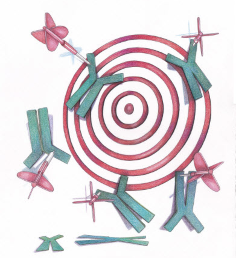
My previous employer grew very quickly from manufacturing licensed clones of common antigens for flow cytometry to eventually undertaking the R&D to develop novel clones across the gamut of biological and technological applications. The kind of validation required for these two efforts are as different as night and day. To clarify, in this instance I’d define common antigens as basic phenotyping markers for cell subsets found in blood. You start the validation having info on the characterizing features of the antigen and its expression. Novel clone development does not always have that benefit. Also, with newly determined antigens or those that were extracted under non-native conditions, often the antibodies we do have for comparison or use as a gold standard are actually not as good as the newly developed ones, leaving us to wonder which result to trust more? And finally, blood is “easy” in that it’s readily accessible, even blood malignancies. Other tissue types, especially if they are human and from a disease state, might be highly inaccessible in abundance and price. Same with validation models that require a specific stimulation where degree of expression or simple human error or other factors that can be out of our control might make the model unreliable. In those cases, when the antibody isn’t working, how can you be sure it’s the antibody and not the model?

Electrophiles and Nucleophiles: Basic Bioconjugation Strategies
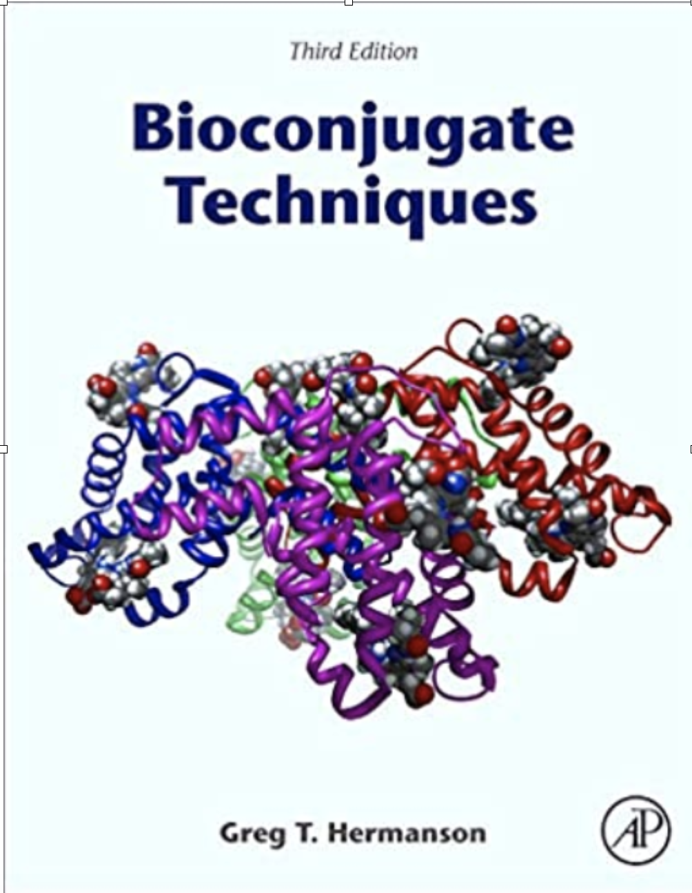
When I began working at Molecular Probes in technical services, my experience was cell biology-centric. The only chemistry knowledge I had back then came from basic courses in university. But, Molecular Probes was at its core a chemical company, albeit fluorescence chemistry. The reagents might be used on cells, but the chemistry was our business. In tech services, our bible was called Bioconjugate Techniques by Greg T. Hermanson.

The Light Inside (literally)
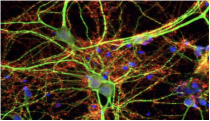
Autofluorescence can be the bane of an experiment, reducing sensitivity in the detection of any emission range especially between 400-550nm. We tend to think of “autofluorescence” as if it were one singular emission source, when in reality it is the consolidation of multiple sources.

The different Personalities of Fluorophores (no one is perfect)
I’ve been lecturing on fluorescent chemistry and chemical probes for almost 20 years (and I’ve also named a lot of those families I complain about)! However, one consistency over the years is how little the average biologist thinks about how their reagents are actually working and thus lumps them all together in one functional basket.
